Skip to content
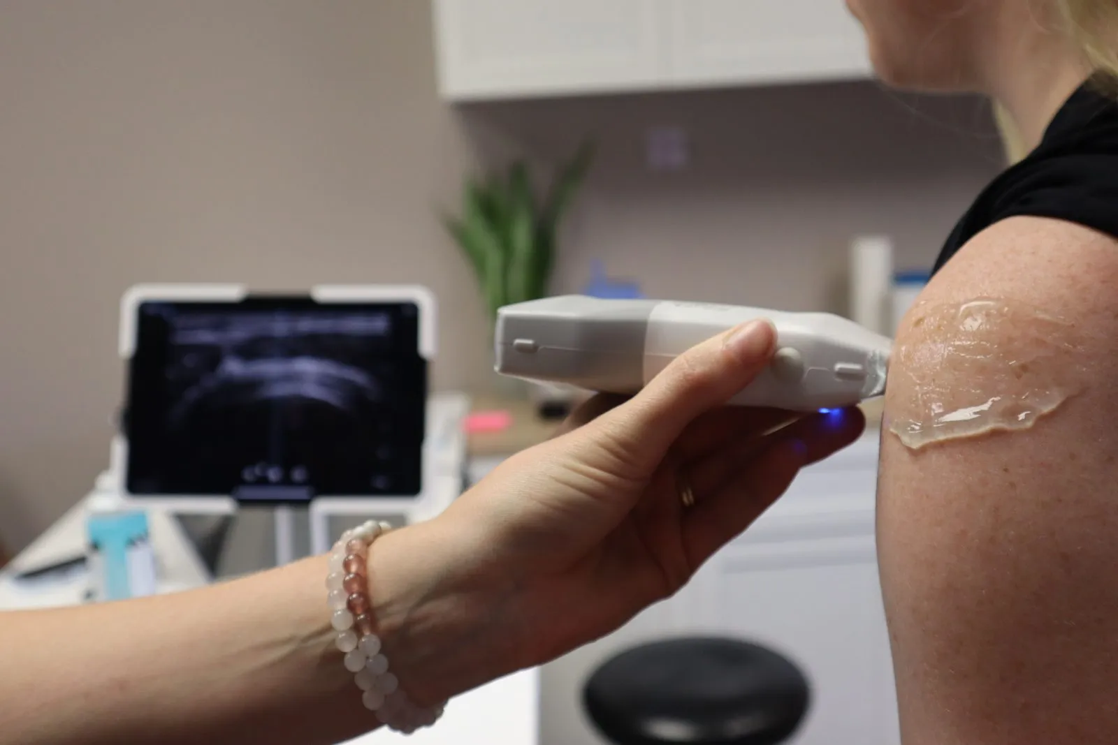











Musculoskeletal ultrasound has become an increasingly useful tool in sports medicine, rheumatology, orthopaedics and other MSK-focused practices.
Its appeal is straightforward: ultrasound provides real-time imaging during the patient encounter and allows clinicians to assess structures dynamically while the patient moves.
But an MSK clinic can ask very different things of an ultrasound system than a general imaging department. The priorities shift, and the most expensive or feature-heavy system is not necessarily the best fit.
Key takeaways
- MSK ultrasound is often incorporated directly into the patient encounter, making workflow, portability and ease of use important considerations.
- A high-frequency linear transducer is the workhorse for many MSK applications and should be one of the first things evaluated when comparing systems.
- Dynamic assessment is one of ultrasound’s major advantages in MSK imaging.
- MRI and ultrasound are complementary modalities. Ultrasound can provide immediate, real-time imaging for appropriate MSK applications without requiring a separate imaging appointment.
- Training matters. MSK ultrasound is highly operator dependent, and the value of a system depends heavily on the people using it.
What is musculoskeletal ultrasound?
Musculoskeletal ultrasound uses diagnostic ultrasound to evaluate structures such as tendons, ligaments, muscles, nerves, bursae and joints.
It can be used for a range of applications, including evaluating soft-tissue injuries, performing dynamic assessments, guiding injections and other procedures, and monitoring certain conditions over time.
One of the characteristics that separates MSK from many other ultrasound applications is that much of the anatomy being examined is relatively superficial. This makes transducer selection particularly important.
It is also commonly used at the point of care. Instead of sending the patient elsewhere for a separate imaging appointment, the clinician may be able to incorporate ultrasound into the visit when appropriate.
Why MSK ultrasound is useful in Canadian clinics
One advantage of MSK ultrasound is the ability to perform imaging at the point of care.
That matters in a Canadian healthcare system where access to diagnostic imaging remains under pressure.
According to the Canadian Institute for Health Information, median wait times increased by 15 days for MRI and 3 days for CT between 2019 and 2024, despite 16% more MRI and CT scans being performed across Canada in 2024 than in 2019.
Ultrasound does not replace MRI or CT. They answer different clinical questions. But for appropriate MSK applications, ultrasound gives clinicians another imaging tool that can be used in the room, in real time, during the patient encounter.
This is part of the broader shift we have discussed in why point-of-care ultrasound is moving into clinics, and MSK is a good example of where that flexibility can be valuable.
What an MSK clinic should look for in an ultrasound system
When we speak with sports medicine and MSK practices, we generally start with the application rather than the machine.
What are you scanning? Who will be scanning? How often will the system be used? Does it stay in one room or move around?
Once those questions are answered, the equipment requirements become much clearer.
1. The right transducer
For many MSK applications, a high-frequency linear transducer is the workhorse.
Higher frequencies provide the detail needed to evaluate superficial anatomy such as tendons, nerves and small joints. But MSK covers a wide range of anatomy and patient types, so there is no single ideal frequency for every examination.
A superficial finger or tendon examination has different requirements than imaging a deeper hip or scanning a larger patient.
Some practices may benefit from a second linear transducer with greater penetration, while others may value a small-footprint transducer for hands, feet and areas where access is limited.
This is why we prefer to start with what you actually plan to scan and work backwards to the transducer and system.
2. Workflow and readiness
In many MSK practices, ultrasound is incorporated into an existing appointment rather than scheduled as a separate imaging study.
That makes workflow important.
How quickly can you get from turning on the system to scanning? How easy is it to select the correct preset? Can you switch transducers quickly? How much adjustment does the image require before you are ready to examine the patient?
These may seem like small details during a product demonstration, but they become much more noticeable when the system is being used repeatedly throughout the day.
When evaluating a system, do not just look at the best image it can produce. Pay attention to how easily you got that image.
3. Dynamic imaging
Dynamic assessment is one of ultrasound’s major advantages in musculoskeletal imaging.
Rather than viewing anatomy only in a static position, ultrasound allows structures to be evaluated in real time while the patient or clinician moves the joint, tendon or surrounding tissue.
That capability can provide information that static imaging cannot provide in the same way.
A 2025 single-centre study published in PeerJ evaluated high-frequency ultrasound in 47 patients with hand trauma and compared the ultrasound findings with surgical findings. The researchers found that incorporating passive motion improved diagnostic performance for several injuries compared with static ultrasound alone.
It is a small study and should not be generalised beyond what the researchers evaluated. But it illustrates an important characteristic of MSK ultrasound: the ability to evaluate anatomy while it moves can be clinically useful.
From an equipment perspective, that means ergonomics matter. The clinician needs to be able to manipulate the transducer, position the patient and operate the system comfortably during a dynamic examination.
4. Portability that matches the practice
“Portable” means different things to different clinics.
Some practices simply need to move the system from one treatment room to another. Others want to take it between locations, onto a field or into another clinical environment.
Those requirements can lead to very different equipment choices.
A compact portable system may make sense when mobility is important. A cart-based system may make more sense when the machine will remain in a dedicated room or when the practice values a larger display, additional transducer capacity, expanded ergonomics or a broader feature set.
The question is not whether portable or cart-based ultrasound is better.
It is where the system needs to go and what it needs to do when it gets there.
For a more detailed comparison, see our guide to cart, portable and handheld ultrasound systems.
What transducer do you need for MSK ultrasound?
This deserves its own discussion because the transducer can have as much impact on an MSK configuration as the system itself.
For many practices, the starting point is a high-frequency linear transducer. From there, the configuration depends on the anatomy and patient population.
A typical conversation might include:
- High-frequency linear: superficial tendons, nerves, muscles and small joints
- Linear with greater penetration: deeper MSK structures and larger patients
- Small-footprint linear: fingers, feet and areas where access is limited
- Additional transducers: selected deeper applications depending on the practice
Buying three probes because they are available does not necessarily make sense.
Neither does choosing a system first and discovering afterwards that the transducer you really need is not available for it.
Choose the applications first. Then build the probe package around them.
Choosing an ultrasound system for an MSK practice
Before comparing individual systems, answer a few practical questions:
- How many ultrasound examinations do you expect to perform in a typical week?
- Will they be scheduled examinations or incorporated into existing appointments?
- Does the system stay in one room, move between rooms or leave the building?
- What anatomy will you scan most often?
- Which transducers do those applications require?
- Who will be scanning and what ultrasound experience do they currently have?
- Do you perform ultrasound-guided injections or other procedures?
- What needs to happen to the images afterwards?
- Do images, measurements or reports need to reach PACS, an EMR or another system?
Those answers can narrow the equipment category considerably before anyone starts comparing model numbers.
That is also why a demonstration is valuable. When possible, scan the types of patients and anatomy you actually see rather than evaluating a system solely from a specification sheet.
Do not overlook training
MSK ultrasound is highly operator dependent.
Transducer position, pressure and angle can all change the appearance of the anatomy being evaluated. Anisotropy in structures such as tendons is a classic example of why scanning technique matters.
That makes training an important part of introducing ultrasound into a practice.
There are also two different types of training to consider.
System and applications training teaches your team how to operate the equipment: controls, transducers, presets, measurements, image optimisation, storage and workflow.
Clinical competency develops through appropriate clinical education, supervised practice and experience.
The two should not be confused.
At Northern Diagnostics, we provide system and applications training with the ultrasound systems we install, using the equipment and configurations your team will actually work with. We train every team we install for, and we remain available for questions that come up after the initial training.
Frequently asked questions
Can musculoskeletal ultrasound replace MRI?
No. Ultrasound and MRI are complementary imaging modalities.
Ultrasound is particularly useful for many superficial soft-tissue structures, dynamic assessment and image-guided procedures. MRI can evaluate anatomy and pathology that ultrasound cannot adequately visualise, including deeper structures and bone marrow.
The appropriate modality depends on the anatomy and clinical question.
What probe do I need for musculoskeletal ultrasound?
For many MSK applications, a high-frequency linear transducer is the starting point because much of the anatomy being examined is relatively superficial.
Some practices benefit from an additional linear transducer with greater penetration for deeper anatomy or larger patients. A small-footprint linear transducer can also be useful for hands, feet and difficult-to-access areas.
The best combination depends on what you actually intend to scan.
Do I need a cart-based system for MSK ultrasound?
Not necessarily.
A portable system can make sense when the ultrasound needs to move between treatment rooms, locations or procedures.
A cart-based system may be preferable when it will remain in a dedicated room or when the practice wants a larger display, additional transducer capacity, different ergonomics or a broader feature set.
The better choice depends on how and where the system will be used.
Is ultrasound useful for guided injections?
Ultrasound can provide real-time visualisation during many image-guided MSK procedures, allowing the clinician to visualise relevant anatomy and the needle during the procedure.
If injections or other guided procedures are an important part of the practice, this should be discussed when choosing the system and transducer. Features such as needle visualisation, ergonomics and the ability to position the display appropriately may become more important.
How long does it take to learn MSK ultrasound?
There is not one timeline.
Previous ultrasound experience, the range of examinations being performed, access to appropriate education and how frequently someone scans all affect the learning process.
Regular practice is important, but purchasing an ultrasound system by itself does not establish clinical competency. Appropriate education and experience remain essential.
Where to start
If you are setting up ultrasound in a sports medicine or MSK practice, the useful first step is not a product comparison.
Start with:
- What do you scan?
- Who will scan it?
- Where will the system be used?
- What needs to happen to the images afterwards?
From there, the appropriate transducers, system category and features become much easier to identify.
At Northern Diagnostics, we are happy to talk through those requirements and help you build a practical configuration around what your clinic actually needs — including when the answer is that you do not need to spend as much as you thought. Get in touch and we will give you an honest view.